Tag Archives: Fluorescence Microscope

With technology evolving by the minute, science in India is advancing beyond par. Microscopy has been a very important branch of research and study and has opened various gates for understanding the cellular structure and micro-organisms. With the fluorescence microscope in India, studies on the cellular level have gotten more advanced and easier.

There are various benefits of bringing Fluorescence microscope in India, some of these benefits are mentioned below.

Easier than standard microscope

The process of using standard microscopes is considered to be a straining process as it takes a lot of time to get good results. Moreover, the study of live-cell tissues through standard microscopy requires one to use several toxic chemicals to get results.

Since the study of live tissues involves pausing the metabolism process in the cell and taking measurements and understanding the cell, a fluorescence microscope works great here.

High-quality results

Another major advantage of going for the Quasmo fluorescence microscope is that it provides high-quality images without consuming much time. This microscope uses filters that remove the background from the image and provide you with a clear cellular picture. In the pictures produced by fluorescence microscopes, you can see every detail of the cell very clearly. You don’t have to be an expert to catch the details by using this microscope.

Non-toxic for specimen

As has been already mentioned, when it comes to live-tissue study, it can be toxic for the specimen. Studying a live tissue is quite difficult since you cannot make measurements and make a record of one particular process. Since the live- cell would have tissues moving or being involved in certain activities, you cannot halt the process and complete your research.

As a result, scientists have to use toxic materials on the specimen to pause the process, take measurements, understand the cell, and then proceed further. Sometimes, the process may lead to the death of the specimen as well. Using a fluorescence microscope eradicates the use of any toxic materials in the process. Since it is highly sensitive and quick, you can make cellular-level measurements in a very short time.

Scope for more complex cell-structure

With fluorescence microscope in India, places like Quasmo has improved our scope to study even more complex and complicated cellular-level problems. Since we get high-quality images in less time and the process is less toxic, scientists are optimistic about understanding the cellular world in even more detail. This will give us more answers about the world and how it works. We may be able to solve fatal diseases as we understand the cell structures to an even greater extent.

To Sum It Up

The fluorescence microscope in India is a revolutionary addition to the field of science as it has opened several opportunities for us to study the world on a cellular level. This type of microscope makes the study work much easier and quicker. It has several benefits which make it a great choice for science to advance even further.

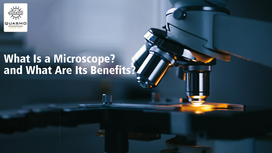
Microscopes are instruments used to view objects that are too small to be seen with the naked eye. They are used in a variety of scientific disciplines, including biology, medicine, and engineering.

What Is a Microscope and What Are Its Uses?

A microscope is an instrument used to magnify objects. It is made of lenses and a light source. A microscope is used to view small objects such as bacteria and other organisms. It is also used to view cells and tissues. There are Quasmo microscopes for biological studies of specimens.

How To Use a Microscope?

A microscope is a device that uses light to magnify objects. There are different types of microscopes, depending on the way the object is magnified. The light microscope uses visible light to magnify an object. The electron microscope uses a beam of electrons to magnify an object.

To use a microscope, you first need to choose the right type of microscope for the type of object you want to magnify. The light microscope is good for magnifying things that are small and thin. The electron microscope is good for magnifying things that are large and thick.

Once the microscope is set up, you need to place the object you want to magnify on the microscope stage. The object should be centered in the microscope lens.

Then you need to adjust the microscope lens to get a good view of the object. You may need to adjust the focus, the brightness, and the contrast of the image.

Once the image is adjusted, you can begin to magnify it. To do this, you need to move the microscope lens up and down. The higher the lens is, the more magnification you will get.
You can also move the microscope lens left and right to get a better view of the object.

To take a picture of the object, you can use the microscope camera. The camera is attached to the microscope and takes pictures of the object through the microscope lens.

What Is a Fluorescent Microscope?

A fluorescent microscope is a microscope that uses fluorescence to image objects. Fluorescence is the emission of light by a substance that has been excited by radiation. In a fluorescent microscope, the object is placed on a slide and a beam of ultraviolet radiation is used to excite the fluorophores on the slide. The fluorophores emit light in all directions and the light is collected by the microscope. The light is then passed through a series of filters that isolate the light emitted by the fluorophores. The image is then projected onto a screen or captured by a camera.

What Are the Benefits of A Fluorescent Microscope?

A fluorescent microscope is a type of microscope that uses fluorescence to increase the contrast of images. This type of microscope has a number of benefits, including the following:

  1. Increased resolution – Fluorescence increases the resolution of images, allowing you to see smaller details. This is because the fluorescent molecules emit light at a specific wavelength, which can be used to create a higher-resolution image.
  2. Increased contrast – The fluorescence of molecules can be used to create a higher contrast image, which makes it easier to see details.
  3. Increased sensitivity – Fluorescence can also be used to increase the sensitivity of a microscope, allowing you to see smaller details in images.
  4. Increased speed – Fluorescence can also be used to increase the speed of a microscope, which can be helpful when you are trying to get a clear image of a fast-moving object.
fluorescence microscope manufacturers in India

For every medical operation or scientific session to be successful, specific equipment needs to be made available to the experts in charge of the operation study. The availability of this equipment along with the required quality is of utmost importance. Amongst the most essential equipment needed in operation lies a microscope of premium quality. All the accuracy required in a medical operation or a study can be provided with a single piece of equipment called a microscope.

Even the tiniest parts of our world, like the microbes, molecules, and other small objects which cannot be seen with the naked eyes, can be well studied using a microscope. Without the availability of microscopes, these things will seem invisible to the human eyes, and no study can be carried-on on the particular object. The efficiency of these microscopes can be well explained with the recent occurrence of the pandemic.

What helped the doctors to diagnose and study the deadly virus of COVID? The short answer to this big question is a microscope. Thousands of lives have only been saved by the availability of microscopes with experts.

The virus’s nature and weaknesses are only known with the perfect study of the virus under a microscope. However, there are microscopes available in the market that serve different purposes. A type of microscope that helped the most in the pandemic is a fluorescent microscope. The fluorescence microscopes in India have proved to be of great help in dealing with the deadly virus of Covid-19. To help you know more about the efficiency of microscopes, we will brief you on the fields in which the microscopes come to their best use.

How are fluorescence microscopes helpful?

1. Investigation with microscopes

Microscopes have come to their best use to scientists and researchers to date. The understanding of the physical and natural world has been made possible with the availability of microscopes to the experts. Microscopes even come to use by scientific researchers in the crime scenes and help them reach out to the clues available at the crime scene. Ultimately, we can say that microscopes can help study the smallest particles of our world.

2. Microscopes in education

The use of microscopes is not limited to scientific researches, but they are also used to teach children about the small structures in the world. Structures that are challenging to be seen with a single human eye can be accurately seen and tested with microscopes.

Final thoughts

We hope that the above information was enough to brief you on the importance of microscopes in the current era. The things that cannot be seen with human eyes can easily be studied using a microscope of premium quality. But if you want to invest in any of the microscopes, let’s say the fluorescence microscope, you are suggested to do the buying from a reputed and well-known fluorescent microscope manufacturer in India. Doing this task responsibly can provide you with long operating lives of the microscopes and get promising outcomes of your investment.

Fluorescence microscope

If we start at the beginning and talk about what a fluorescence microscope is and what it does, we can say that it is an optical microscope that studies the properties of organic and non-organic objects using fluorescence and phosphorescence instead of or in addition to reflection or absorption.

The light emitted by a substance with absorbed light or another electromagnetic radiation is known as fluorescence.

When we discuss what phosphorescence is, we can say that it is a sort of photo-luminescence similar to fluorescence.

In the early twentieth century, the fluorescence microscope was invented. This incredible invention is performing admirably.

Fluorescence microscopy is a powerful analytical tool that combines light microscopy’s magnifying properties with fluorescence visibility. It is a phenomenon in which a fluorescent molecule called a fluoroscope absorbs and emits a small range of light wavelengths. This microscopy is performed by combining a bright light source, specific filters, and a method of fluorescent marking material with a basic light microscope.

What is the underlying principle?

This procedure is based on a simple idea. When light reaches the arc lamp, it is directed towards the exciter filter, which determines the excavation length.

Structure and Working

The fluorescent microscope’s principal components are quite similar to those of a standard light microscope. The type of light source and the employment of specific filter components are the two key variations.

The light source lamp, excitation filter, dichroic mirror, and emission filter are all standard components of this microscope. Getting this top-notch source from the leading microscope manufacturer in Ambala is a piece of cake.

Through the objective lens, the light of the excitation wavelength is focused on the specimen. The objective focuses on the fluorescence emitted by the specimen on the detector. Only reflected excitation light and the emitted light reach the aim since most of the excitation light is transmitted through the sample.

What Are The Applications of This Microscope?

This microscope is handy for spotting features in both preserved and living biological samples.

Today, this microscope is standard equipment for scientific research and processes, and this incredible discovery is changing the face of science. The fluorescence microscope in India has been steadily expanding in popularity owing to the growing demand for this technology. The reason for its ever-increasing magnitude is the quality of service provided by this source. It allows for multi color staining, structural labeling, and the measurement of a cell’s physiological condition.

Advantages

Fluorescence microscopy is one of the most popular technologies for live-cell observation and elucidating the structure of bio molecules in tissues and cells. It allows them to be studied without the need for hazardous and time-consuming staining processes. Cell dynamics can be examined in living samples with a precision and sensitivity capable of tracking the path of a single protein throughout its lifetime, or samples can be fixed before the addition of a fluoroscope, halting the metabolism of cells at a point in time and allowing detailed measurements of the preserved specimen.

When employed in conjunction with con focal microscopy, the great degree of specificity provided by contemporary fluoroscope probes allows specific proteins or other biological components to be stressed and highlighted. This results in a clear three-dimensional internal structure of the cell. Multiple overlapping systems can be discovered simultaneously within the same sample due to the diversity and specificity of the available probes.


Fluorescence Microscope In India

Fluorescence microscopy is an essential tool for studying cell physiology. Fluorescence microscopy is a type of microscope that studies the properties of organic and inorganic materials by using fluorescence and phosphorescence.

Fluorescence is the emission of light by a substance that has absorbed light or other electromagnetic energy while phosphorescence is a kind of optical property that is related to fluorescence.

Microscopy Is a Basic Research Tool

Microscopes are significant research and discovery tools that have helped in the discovery of numerous ground-breaking discoveries over the years. Fluorescence microscopy integrates the magnifying properties of a light microscope with fluorescence technology, which allows the activation of fluorophores – fluorescent chemical substances and the measurement of their emissions.

Fluorescence microscope in India allows scientists to observe where specific cell types or chemicals are present inside tissues or cells.

Components of Fluorescence Microscopy

  • Fluorescent dyes (Fluorophore)
  • A light source
  • The excitation filter
  • The dichroic mirror

Applications:

Fluorescence microscopy is used to detect various structures, chemicals, or proteins inside the cell. Almost every component of living or fixed cells and tissues can be labelled and examined using the technique at high magnifications.

When it comes to fluorescence microscopy, the fluorophore can be just as important as the microscope itself, and the type of fluorophore photographed affects the excitation wavelength and detection wavelength. The excitation wavelengths contain a small range of energy that the fluorophore can absorb and cause it to transition to an active state.

Once stimulated, a wide range of excited, a variety of emissions or transitions back to a lower energy state is prepared, leading to an emission spectrum.

Fluorescence Microscopy for Cell Labeling

Fluorophores, such as fluorescent dyes, are used to label cell structures, significantly increasing the visible qualities of biological or chemical structures. These can be modified or combined to have specific target ability in medical applications. High-specificity fluorophores are commonly used in medical diagnostics, and new research is finding new applications for them.

Fluorescence Microscopy for Protein Characterization

Fluorescence microscopy can be used in a variety of experiments that use different types of fluorophores. The detection of proteins that have been labeled with antibodies that are attached to or “conjugated” to fluorescent compounds is one of the most common applications of fluorescence microscopy. Intracellular dynamic protein interactions are important for a wide range of biochemical functions.

 We are the best fit for you if you are looking for Fluorescence Microscopy in India

Quasmo India Microscope trades in several different microscopes. We offer Fluorescent Research Microscopes PZQ-102, PZQ-106, and Fluorescent Research Microscopes QX-50, Star- 7FL, and Malaria Detection Microscope PZQ-101 that are the best categories of our Fluorescence Microscopy in India.

In the Ultraviolet wavelength range, we provide a variety of continuously variable filters capable of examining the spectral properties of a specific sample with exceptional transmission values and deep blocking. If you would like any more information about our products, you can contact us, we always available to assist you.